Hip Anatomy
Hip Joint
The hip joint is the largest weight-bearing joint in the human body. It is also referred to as a ball and socket joint and is surrounded by muscles, ligaments, and tendons. The thigh bone or femur and the pelvis join to form the hip joint.
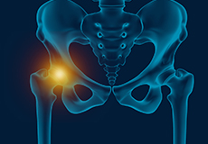
Any injury or disease of the hip will adversely affect the joint's range of motion and ability to bear weight.
The hip joint is made up of the following:
- Bones and joints
- Ligaments of the joint capsule
- Muscles and tendons
- Nerves and blood vessels that supply the bones and muscles of the hip
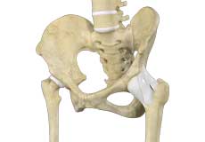
Bones and Joints
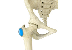
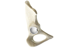
The hip joint is the junction where the hip joins the leg to the trunk of the body. It is comprised of two bones: the thigh bone or femur and the pelvis which is made up of three bones called ilium, ischium, and pubis. The ball of the hip joint is made by the femoral head while the socket is formed by the acetabulum. The Acetabulum is a deep, circular socket formed on the outer edge of the pelvis by the union of three bones: ilium, ischium, and pubis. The lower part of the ilium is attached by the pubis while the ischium is considerably behind the pubis. The stability of the hip is provided by the joint capsule or acetabulum and the muscles and ligaments which surround and support the hip joint.
The head of the femur rotates and glides within the acetabulum. A fibrocartilagenous lining called the labrum is attached to the acetabulum and further increases the depth of the socket.
The femur or thigh bone is one of the longest bones in the human body. The upper part of the thigh bone consists of the femoral head, femoral neck, and greater and lesser trochanters. The head of the femur joins the pelvis (acetabulum) to form the hip joint. Next, to the femoral neck, there are two protrusions known as greater and lesser trochanters which serve as sites of muscle attachment.
Articular cartilage is the thin, tough, flexible, and slippery surface lubricated by synovial fluid that covers the weight-bearing bones of the body. It enables smooth movements of the bones and reduces friction.
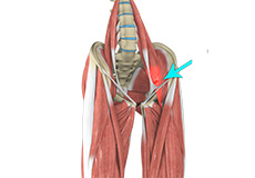
Ligaments
Ligaments are fibrous structures that connect bones to other bones. The hip joint is encircled with ligaments to provide stability to the hip by forming a dense and fibrous structure around the joint capsule. The ligaments adjoining the hip joint include:
- Iliofemoral ligament: This is a Y-shaped ligament that connects the pelvis to the femoral head at the front of the joint. It helps in limiting the over-extension of the hip.
- Pubofemoral ligament: This is a triangular shaped ligament that extends between the upper portion of the pubis and the iliofemoral ligament. It attaches the pubis to the femoral head.
- Ischiofemoral ligament: This is a group of strong fibers that arise from the ischium behind the acetabulum and merge with the fibers of the joint capsule.
- Ligamentum teres: This is a small ligament that extends from the tip of the femoral head to the acetabulum. Although it has no role in hip movement, it does have a small artery within that supplies blood to a part of the femoral head.
- Acetabular labrum: The labrum is a fibrous cartilage ring which lines the acetabular socket. It deepens the cavity, increasing the stability and strength of the hip joint.
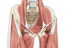
Muscles and Tendons
A long tendon called the iliotibial band runs along the femur from the hip to the knee and serves as an attachment site for several hip muscles including the following:
- Gluteals: These are the muscles that form the buttocks. There are three muscles (gluteus minimus, gluteus maximus, and gluteus medius) that attach to the back of the pelvis and insert into the greater trochanter of the femur.
- Adductors: These muscles are located in the thigh which helps in adduction, the action of pulling the leg back towards the midline.
- Iliopsoas: This muscle is located in front of the hip joint and provides flexion. It is a deep muscle that originates from the lower back and pelvis and extends up to the inside surface of the upper part of the femur.
- Rectus femoris: This is the largest band of muscles located in front of the thigh. They also are hip flexors.
- Hamstring muscles: These begin at the bottom of the pelvis and run down the back of the thigh. Because they cross the back of the hip joint, they help in extension of the hip by pulling it backward.
Nerves and Arteries
Nerves of the hip transfer signals from the brain to the muscles to aid in hip movement. They also carry the sensory signals such as touch, pain, and temperature back to the brain.
The main nerves in the hip region include the femoral nerve in the front of the femur and the sciatic nerve at the back. The hip is also supplied by a smaller nerve known as the obturator nerve.
In addition to these nerves, there are blood vessels that supply blood to the lower limbs. The femoral artery, one of the largest arteries in the body, arises deep in the pelvis and can be felt in front of the upper thigh.
Hip Movements
All of the anatomical parts of the hip work together to enable various hip movements. Hip movements include flexion, extension, abduction, adduction, circumduction, and hip rotation.
Hip Conditions
Snapping Hip Syndrome
Snapping hip syndrome is named for the sound or feel of a catching tendon across the hip joint. It is most common in gymnasts, soccer players, runners and rowers between the ages of 15-35 years old. There are two main types: Internal and External.

Athletic Pubalgia
Athletic pubalgia, also known as sports-hernia and Gilmore’s groin, is a condition characterized by chronic pain due to damage or strain to the soft tissues in the groin and pelvic region. This condition is mostly observed in athletes undergoing vigorous movements where there is twisting or a sudden change in direction. Although this condition is not a true hernia, it can lead to one.
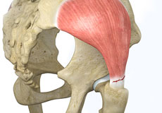
Hip Abductor Tears
Hip abductors are a major group of muscles found in the buttocks. It includes the gluteus maximus, gluteus medius, gluteus minimus, and tensor fascia lata muscles.
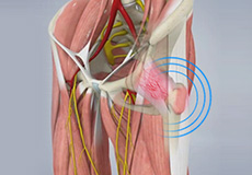
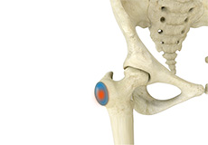
Hip Bursitis
Hip bursitis is a painful condition caused by the inflammation of a bursa in the hip. Bursae are fluid-filled sacs present in the joints between bone and soft tissue to reduce friction and provide cushioning during movement.
Hip Labral Tear
A hip labral tear is an injury to the labrum, the cartilage that surrounds the outside rim of your hip joint socket.
Hip Ligament Injuries
Injuries to the hip ligaments are commonly called a hip sprain and can range from minor tears of the ligaments to more serious injuries involving the hip muscles, tendons or bone.

Osteoarthritis of the Hip
Osteoarthritis, also called degenerative joint disease, is the most common form of arthritis. It occurs most often in the elderly. This disease affects the tissue covering the ends of bones in a joint called cartilage. In osteoarthritis, the cartilage becomes damaged and worn out, causing pain, swelling, stiffness and restricted movement in the affected joint. Although osteoarthritis may affect various joints including the hips, knees, hands, and spine, the hip joint is most commonly affected. Rarely, the disease may affect the shoulders, wrists, and feet.
Hip Tendonitis
Tendons are strong connective tissue structures that connect muscle to bone. Hip tendonitis is a condition associated with degeneration of the hip tendons. This condition is mainly caused due to strain on the tendons which may occur due to overuse or biomechanical problems.
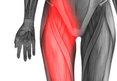
Hip Flexor Strain
A hip flexor strain is an overuse injury to the flexor muscles of your hip and can range from a minor stretch injury to a complete tear of the muscle fibers or tendons.
Trochanteric Bursitis
Trochanteric bursitis is a painful condition caused by inflammation of the trochanteric bursa, a fluid-filled sac that overlies the greater trochanter (bony prominence at the outer side of the hip). Bursae are present at various regions of the body between the bones and tendons to reduce friction with movement.
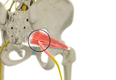
Iliopsoas Tendonitis
Iliopsoas tendonitis also referred to as snapping hip syndrome, is an inflammation of the iliopsoas tendon or the surrounding area. The iliopsoas is the hip flexor tendon located over the front of the hip socket. The term snapping hip describes the sound made, a snap or click, that occurs with certain hip movements including flexion, extension, and rotation of the hip.
Hip Flexor Pain
Hip flexor pain is a distressing feeling or discomfort noted in the hip and/or groin region that can make everyday activities, such as going up and down the stairs or lifting your leg to tie a shoe extremely difficult and painful and can severely limit your activity and mobility.
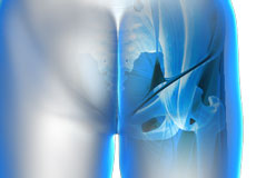
Deep Gluteal Pain Syndrome
Deep gluteal pain syndrome is a medical condition characterized by severe pain, numbness, or tingling in the buttock or hip that may radiate down the back of the leg. It occurs due to the compression of nerves such as the sciatic nerve and the pudendal nerve in the gluteal (buttock) region. Deep gluteal pain syndrome can include a group of conditions such as sciatica and piriformis syndrome which cause similar symptoms.
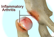
Inflammatory Arthritis of the Hip
The inflammation of the joints is referred to as arthritis. Inflammation arises when the smooth lining called cartilage at the ends of bones wears away. In some cases, the inflammation is caused when the lining of the joint becomes inflamed as part of an underlying systemic disease. These conditions are referred to as inflammatory arthritis.
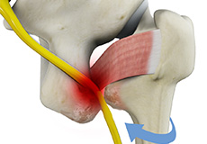
Ischiofemoral Impingement
Ischiofemoral impingement is a condition in which there is an abnormal contact (impingement) between the soft tissues of the thigh bone and hip due to the narrowing of space between the lesser trochanter and ischium, resulting in significant hip pain.
Hip Procedures
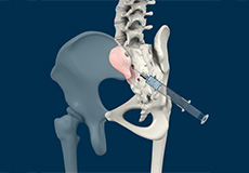
Sacroiliac Joint Injections
Any inflammation or irritation in SI joints may cause pain in the lower back, abdomen, groin, buttocks or legs. Sacroiliac joint injections can be used both for diagnostic as well as therapeutic purposes for pain. As a diagnostic tool, it helps your doctor locate the origin of pain. For therapeutic uses, SI joint injections will contain a steroid medication along with an anesthetic agent to provide relief from pain for a longer duration.
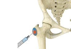
Trochanteric Bursa Injections
A trochanteric bursa injection is a minimally invasive procedure in which medicine is injected directly into the trochanteric bursa in the hip joint using a thin needle and syringe to relieve pain and inflammation. The injection usually contains a combination of numbing medicine and cortisone (an anti-inflammatory agent). Trochanteric bursitis, also known as greater trochanteric bursitis or hip bursitis, is the main indication for a trochanteric bursa injection.
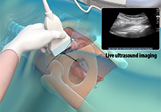
Ultrasound Guided Hip Injections
An ultrasound scan is an imaging procedure that uses high-frequency sound waves to produce pictures of the inside of the body. Ultrasound-guided hip joint injections are used to diagnose the underlying cause and relieve hip pain. The injection consists of a special mixture of an anesthetic and a steroid that blocks pain impulses and reduces inflammation in the injected area.